Learn about the Biomedical technology
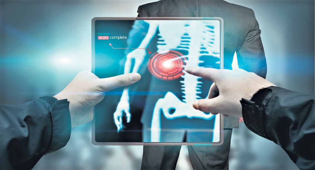
Hyderabad: This article is in continuation of previous articles that talk about the biodiversity, environment and ecology. Here we would be focusing on Biomedical technology. Biomedical Technology Biomedical technology is the application of principles of biophysics and biochemistry for studying certain biological aspects. It involves: a. X-ray imaging/X-ray Radiography b. CAT/CT scan (Computed Axial Tomography/Computed […]
Hyderabad: This article is in continuation of previous articles that talk about the biodiversity, environment and ecology. Here we would be focusing on Biomedical technology.
Biomedical Technology
Biomedical technology is the application of principles of biophysics and biochemistry for studying certain biological aspects. It involves:
a. X-ray imaging/X-ray Radiography
b. CAT/CT scan (Computed Axial Tomography/Computed Tomography)
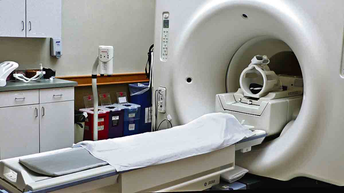
c. MRI Scan (Magnetic Resonance Imaging)
d. PET Scan (Positron Emission Tomography)
e. Sonography
f. ECG (Electrocardiogram/Electrocardiograph)
g. EEG (Electroencephalography)
h. ELISA (Enzyme-linked immunosorbent assay)
i. Western blot technique, etc.
X- ray Radiography
- X-ray discovered by Rontgen
- X-ray are useful in diagnostic radiography.
- It is a simply radiological technique to observe the human body parts & diagnose any physical (anatomical) variations.
- X-ray images show areas of different densities and composition in different identifiable/diagnosable forms.
- A beam of X-rays is produced by an X-ray generator and is projected on the body parts.
- X-rays that pass through the body parts are recorded on a photographic film/ observed on a fluorescent screen.
- Photographs developed using x-rays are known as radiographs/skiagraphs.
- Dense bones absorb much of the x-radiation and soft tissues allow more X-rays to pass through.
- So, bones appear more white and soft tissues look greyish calcifications in tissues to appear whitish air in the lungs appears blackish as it does not block.
X-rays
- The X-ray film provides a ‘2D’ representation of all the structures of the body part superimposed on each other (X-ray radiography)/ it can be stored as a digital image.
- It is used to diagnose/ treat patients by assessing the presence absence of disease, foreign objects, and structural damage/ other anomalies.
- Angiography involves taking a series of x-rays as blood flows through blood vessels such as the coronary/ carotid arteries to study blocks in them.
- This procedure involves injection of dyes in to blood to get contrast image and now a days digital subtraction angiography is used to get a better resolution images.
- Portable X-ray machines are also available, nowadays.
- X-rays help to detect skeletal fractures, pneumonia, tuberculosis, cancer etc.
- Every precaution should be taken to avoid exposure of gonds and foetus of pregnant women, as X-rays may prove to be more dangerous to them.
Computed Axial Tomography (CAT)
OR
Computed Tomography (CT)
- Computed/ computerized axial tomography (CAT) or computed tomography (CT) Scan is a medical imaging method that employees tomography (a process of producing a two dimensional slice through a 3 dimensional object.
- A large donut shaped X-ray machine takes X-ray images at many different angles around the body.
- The X-ray detector of the CT-Scanner can detect hundreds of different levels of density of tissues in body organs.
- The data is transmitted to a computer which builds up 3-D cross sectional picture of the part of the body and displays the picture on the screen. This recorded image is called tomogram.
- The body can be seen on CAT/CT scan as slices from the skin to the central part.
- A CAT scanner emits a series of narrow beams of X-rays through the human body as it moves through an arc, unlike an X-ray machine which sends just one radiation beam.
- The final 3-D picture of a CAT scan is far more superior and detailed than an ordinary X-ray picture, sometimes; patients may be given a contrast dye for better resolution.
- CAT scans are useful to measure accurately the density of bones in evaluating Osteoporosis.
- CAT scans are performed to analyse the head injuries (blood clots and skull fractures) to know the anatomy of various visceral organs.
Dr. Modala Mallesh
Subject Expert
Palem, Nakrekal, Nalgonda
9989535675