Novel microscopy method provides look into future of cell biology
The invention, described in an article in the journal Nature Methods, could open new avenues in advanced microscopy, the researchers say.

Washington: UT (University of Texas) Southwestern scientists collaborated with colleagues in England and Australia to build and test a novel optical device that converts commonly used microscopes into multiangle projection imaging systems.
The invention, described in an article in the journal Nature Methods, could open new avenues in advanced microscopy, the researchers say.
“It is a completely new technology, although the theoretical foundations for it can be found in old computer science literature,” said corresponding author Reto Fiolka, PhD. Both he and co-author Kevin Dean, PhD, are assistant professors of cell biology and in the Lyda Hill Department of Bioinformatics at UT Southwestern.
“It is as if you are holding the biological specimen with your hand, rotating it, and inspecting it, which is an incredibly intuitive way to interact with a sample. By rapidly imaging the sample from two different perspectives, we can interactively visualize the sample in virtual reality on the fly,” said Dean, director of the UTSW Microscopy Innovation Laboratory, which collaborates with researchers across campus to develop custom instruments that leverage advances in light microscopy.
Currently, acquiring 3D-image information from a microscope requires a data-intensive process, in which hundreds of 2D images of the specimen are assembled into a so-called image stack.
To visualize the data, the image stack is then loaded into a graphics software program that performs computations to form two-dimensional projections from different viewing perspectives on a computer screen, the researchers explain.
“Those two steps require a lot of time and may need a very powerful and expensive computer to interact with the data,” Fiolka said.
The team realized it could form projections from multiple angles by optical means, bypassing the need to acquire image stacks and rendering them with a computer. This is achieved by a simple and cost-effective unit consisting of two rotating mirrors that are inserted in front of the camera of the microscope system.
“As a result, we can do all this in real-time, without any noticeable delay. Surprisingly, we can look from different angles ‘live’ at our samples without rotating the samples or the microscope,” Fiolka said. “We believe this invention may represent a new paradigm for acquiring 3D information via the fluorescence microscope.”
It also promises incredibly fast imaging. While an entire 3D-image stack may require hundreds of camera frames, the new method requires only one camera exposure.
Initially, the researchers developed the system with two common light-sheet microscopes that require a post-processing step to make sense of the data. That step is called de-skewing and essentially means rearranging the individual images to remove some distortions of the 3D-image stack. The scientists originally sought to perform this de-skewing optically.
While experimenting with the optical de-skewing method, they realized that when they used an incorrect amount of “de-skew,” the projected image seemed to rotate.
“This was the aha! moment. We realized that this could be bigger than just an optical de-skewing method; that the system could work for other kinds of microscopes as well,” Fiolka said.
“This study confirms the concept is more general,” Dean said. “We have now applied it to various microscopes, including light-sheet and spinning disk confocal microscopy.”
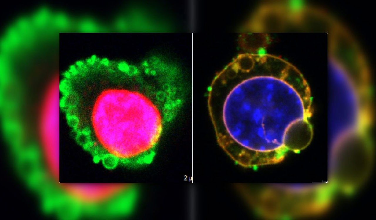
Using the new microscope method, they imaged calcium ions carrying signals between nerve cells in a culture dish and looked at the vasculature of a zebrafish embryo. They also rapidly imaged cancer cells in motion and a beating zebrafish heart.