Do you know your digestive system well?
By Dr Modala Mallesh, Subject Expert Palem, Nakrekal, Nalgonda Ph. 9989535675 This article focuses on the digestive system and absorption of food in the body. Aspirants can expect questions on some basic biological workings of a human body in the upcoming competitive examinations. DIGESTION AND ABSORPTION • Food is one of the basic requirements of […]

By Dr Modala Mallesh,
Subject Expert
Palem, Nakrekal,
Nalgonda
Ph. 9989535675
This article focuses on the digestive system and absorption of food in the body. Aspirants can expect questions on some basic biological workings of a human body in the upcoming competitive examinations.
DIGESTION AND ABSORPTION
• Food is one of the basic requirements of all living organisms.
• The major components of our food are carbohydrates, proteins and fats. Vitamins and minerals are also required in small quantities.
• Food provides energy and organic materials for growth and repair of tissues. The water we take in, plays an important role in metabolic processes and also prevents dehydration of the body.
• Biomacromolecules in food cannot be utilised by our body in their original form.
• They have to be broken down and converted into simple substances in the digestive system.
• This process of conversion of complex food substances to simple absorbable forms is called digestion and is carried out by our digestive system by mechanical and biochemical methods.
DIGESTIVE SYSTEM
• The human digestive system consists of the alimentary canal and the associated glands (Digestive Glands).
ALIMENTARY CANAL
• The alimentary canal begins with an anterior opening – the mouth, and it opens out posteriorly through the anus.
• The mouth leads to the buccal cavity or oral cavity.
• The oral cavity has a number of teeth and a muscular tongue.
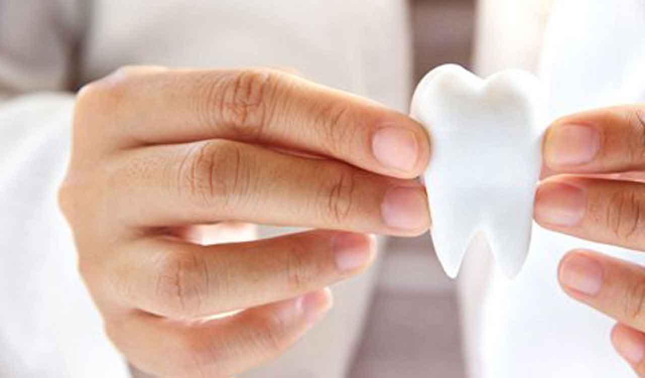
Teeth
• Each tooth is embedded in a socket of jaw bone. This type of attachment is called thecodont.
• Majority of mammals including human being forms two sets of teeth during their life, a set of temporary milk or deciduous teeth replaced by a set of permanent or adult teeth. This type of dentition is called diphyodont.
• An adult human has 32 permanent teeth which are of four different types (Heterodont dentition), namely, incisors (I), canine (C), premolars (PM) and molars (M).
• Arrangement of teeth in each half of the upper and lower jaw in the order I, C, PM, M is represented by a dental formula which in human is 2123/2123.
• The hard chewing surface of the teeth, made up of enamel, helps in the mastication of food.
The Tongue
• The tongue is a freely movable muscular organ attached to the floor of the oral cavity by the frenulum.
• The upper surface of the tongue has small projections called papillae, some of which bear taste buds.
• The oral cavity leads into a short pharynx which serves as a common passage for food and air.
• The oesophagus and the trachea (wind pipe) open into the pharynx.
• A cartilaginous flap called epiglottis prevents the entry of food into the glottis – opening of the wind pipe – during swallowing.
• The oesophagus is a thin, long tube which extends posteriorly passing through the neck, thorax and diaphragm and leads to a ‘J’ shaped baglike structure called stomach.
• A muscular sphincter (gastro-oesophageal) regulates the opening of oesophagus into the stomach.
• The stomach, located in the upper left portion of the abdominal cavity, has 3 major parts – a cardiac portion into which the oesophagus opens, a fundic region, body (main central region) and a pyloric portion which opens into the first part of small intestine.
• Small intestine is distinguishable into three regions – a ‘C’ shaped duodenum, a long coiled middle portion jejunum and a highly coiled ileum.
• The opening of the stomach into the duodenum is guarded by the pyloric sphincter.
• Ileum opens into the large intestine.
• Large intestine consists of caecum, colon and rectum.
• Caecum is a small blind sac which hosts some symbiotic micro-organisms.
• A narrow finger-like tubular projection, the vermiform appendix which is a vestigial organ, arises from the caecum.
• The caecum opens into the colon.
• The colon is divided into four parts – an ascending, a transverse, descending part and a sigmoid colon.
• The descending part opens into the rectum which opens out through the anus.
• The wall of alimentary canal from oesophagus to rectum possesses four layers namely serosa, muscularis, sub-mucosa and mucosa.
• Serosa is the outermost layer and is made up of a thin mesothelium (epithelium of visceral organs) with some connective tissues.
• Muscularis is formed by smooth muscles usually arranged into an inner circular and an outer longitudinal layer. An oblique muscle layer may be present in some regions.
• The sub-mucosal layer is formed of loose connective tissues containing nerves, blood and lymph vessels. In duodenum, glands are also present in sub-mucosa.
• The innermost layer lining the lumen of the alimentary canal is the mucosa. This layer forms irregular folds (rugae) in the stomach and small finger-like foldings called villi in the small intestine.
• The cells lining the villi produce numerous microscopic projections called microvilli giving a brush border appearance. These modifications increase the surface area enormously. Villi are supplied with a network of capillaries and a large lymph vessel called the lacteal.
• Mucosal epithelium has goblet cells which secrete mucus that help in lubrication. Mucosa also forms glands in the stomach (gastric glands) and crypts in between the bases of villi in the intestine (crypts of Lieberkuhn).
• All the four layers show modifications in different parts of the alimentary canal.