Know all about muscle structure
Hyderabad: This article is in continuation to the previous articles focusing on Locomotion and Movement. Today, we’ll examine the skeletal muscle in detail. Visceral muscles • Visceral muscles are located in the inner walls of hollow visceral organs of the body like the alimentary canal, reproductive tract, etc. • They do not exhibit any striation […]
Hyderabad: This article is in continuation to the previous articles focusing on Locomotion and Movement. Today, we’ll examine the skeletal muscle in detail.
Visceral muscles
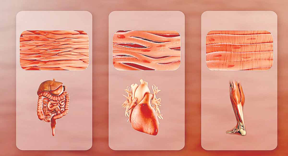
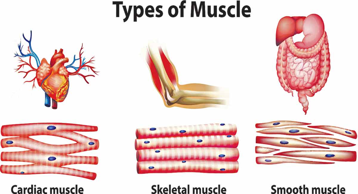
• Visceral muscles are located in the inner walls of hollow visceral organs of the body like the alimentary canal, reproductive tract, etc.
• They do not exhibit any striation and are smooth in appearance. Hence, they are called smooth muscles (nonstriated muscle).
• Their activities are not under the voluntary control of the nervous system and are therefore known as involuntary muscles.
• They assist, for example, in the transportation of food through the digestive tract and gametes through the genital tract.
Skeletal muscle
• Let us examine a skeletal muscle in detail to understand the structure and mechanism of contraction.
• Each organised skeletal muscle in our body is made of a number of muscle bundles or fascicles held together by a common collagenous connective tissue layer called fascia.
• Each muscle bundle contains a number of muscle fibres.
• Each muscle fibre is lined by the plasma membrane called sarcolemma enclosing the sarcoplasm.
• Muscle fibre is a syncytium as the sarcoplasm contains many nuclei.
• The endoplasmic reticulum, i.e., sarcoplasmic reticulum of the muscle fibres is the store house of calcium ions.
Myofilaments or Myofibrils
• A characteristic feature of the muscle fibre is the presence of a large number of parallelly arranged filaments in the sarcoplasm called myofilaments or myofibrils.
• Each myofibril has alternate dark and light bands on it.
• A detailed study of the myofibril has established that the striated appearance is due to the distribution pattern of two important proteins – Actin and Myosin.
• The light bands contain actin and is called I-band or Isotropic band, whereas the dark band called ‘A’ or Anisotropic band contains myosin.
• Both the proteins are arranged as rod-like structures, parallel to each other and also to the longitudinal axis of the myofibrils.
• Actin filaments are thinner as compared to the myosin filaments, hence are commonly called thin and thick filaments respectively.
• In the centre of each ‘I’ band is an elastic fibre called ‘Z’ line which bisects it.
• The thin filaments are firmly attached to the ‘Z’ line.
• The thick filaments in the ‘A’ band are also held together in the middle of this band by a thin fibrous membrane called ‘M’ line.
• The ‘A’ and ‘I’ bands are arranged alternately throughout the length of the myofibrils.
• The portion of the myofibril between two successive ‘Z’ lines is considered as the functional unit of contraction and is called a sarcomere.
• In a resting state, the edges of thin filaments on either side of the thick filaments partially overlap the free ends of the thick filaments leaving the central part of the thick filaments.
• This central part of thick filament, not overlapped by thin filaments is called the ‘H’ zone.
By
Dr. Modala Mallesh
Subject Expert
Palem, Nakrekal, Nalgonda
9989535675