Microscopic pictures that won at the Nikon Small World competition
From the colourful head of a tick to the scales on a butterfly, these microscopic pictures reveal an unnoticed world
Hyderabad: From the smallest creatures to the biggest ones, the microscopic view is way intriguing and different from what is seen with the naked eye. Having realised this, Jason Kirk, director of a microscope-imaging facility at Baylor College of Medicine, started to tinker with microscopes and used them to photograph several things from his backyard.
Having chosen a Southern live Oak tree, Kirk illuminated the leaf’s small-scale structures with a customised microscope. He then captured about 200 images and stacked them on top of each other to create this vibrant portrait. Have a look:
The result shows the leaf’s trichomes – little outgrowths that protect the plant – in white. Pores that help the plant regulate gas flows, called stomata, appear in purple. Vessels that carry water through the leaf pop out in blue-green, according to ‘Business Insider’.
That technical handiwork led Kirk to win first place in this year’s Nikon Small World competition – a contest launched in 1974 by the camera company – to recognise the achievements in microscope photography, also known as microscopy. The Nikon Small World competition was created to show the world how art and science come together under the microscope.
For the 2021 competition, Nikon received nearly 1,900 entries from 88 countries. A panel of judges selected 20 winners and recognised 80 other images for distinction or honourable mention. Here are some of the pictures that made it big at the competition:
A microfluidic device
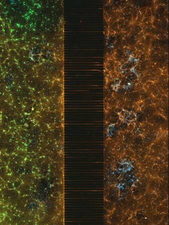
A microfluidic device containing 300k networking neurons in two isolated populations! Both sides were treated with a unique virus and bridged by axons. The picture won second place in the competition.
Photograph: Esmeralda Paric and Holly Stefen/ Dementia Research Centre, Macquarie University
A louse
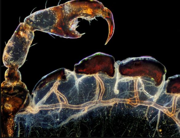
The rear leg, claw and respiratory trachea of a louse (Haematopinus suis).
Photograph: Frank Reiser/ Nassau Community College
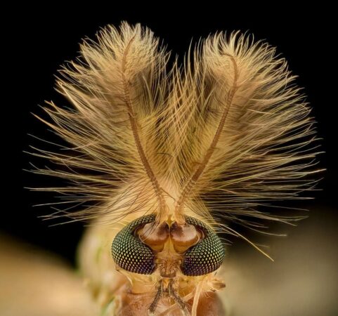
The head of a tick
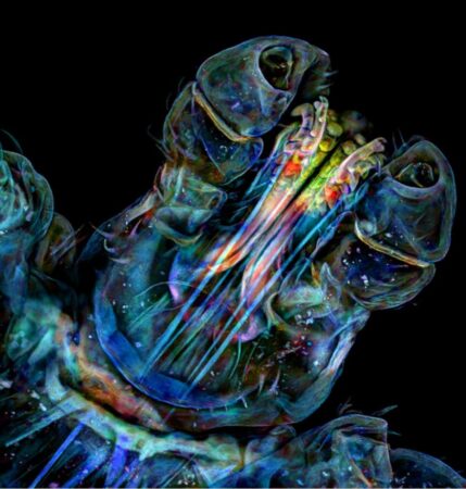
A microscopic view of the head of a tick reveals some hidden colours.
Photograph: Dr Tong Zhang and Dr Paul Stoodley/ The Ohio State University
Tiny insect
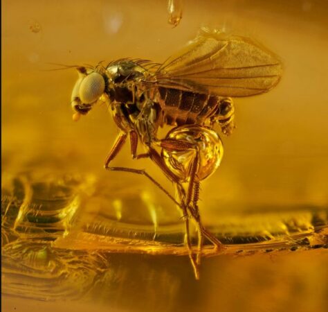
An amber series project displays tiny insects no more than 3mm long that have been encapsulated with hardened tree sap for 45 million years.
Photograph: Levon Bliss Photography Ltd.
Butterfly wing
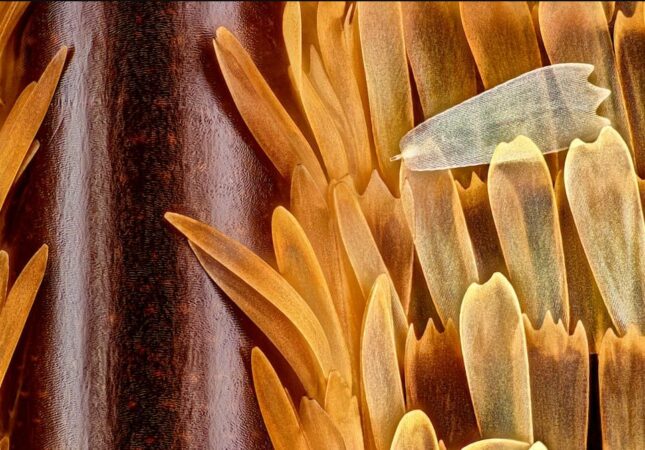
Vein and scales on a butterfly wing (Morpho didius).
Photograph: Sebastien Malo
Cotton fabric with pollen grains
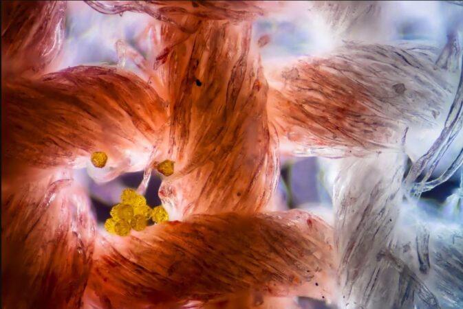
The microscopic picture captures how the fibres weave together. The yellow circles are a few stray grains of pollen that got caught.
Photograph: Felice Placenti
A midge
A midge (Chironomidae diptera) photographed by a customised microscope.
Photograph: Dr Erick Francisco Mesen
A Housefly
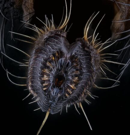
The proboscis of a housefly (Musca domestica).
Photograph: Oliver Dum
Hatched brine shrimp eggs
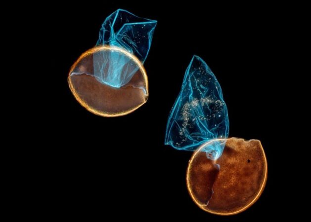
Two hatched brine shrimp eggs after the babies had hatched (darkfield, fluorescence, image stacking).
Photograph: Waldo Nell
Now you can get handpicked stories from Telangana Today on Telegram everyday. Click the link to subscribe.
Click to follow Telangana Today Facebook page and Twitter .